Recent insight into poultry trace mineral nutrition

Poultry Focus Asia, janvier 2018

Trace mineral nutrition is in the spotlight. Covering the nutritional needs of high producing animals is essential to achieve best performances. Trace elements like copper, zinc, manganese and selenium are essential for critical body functions and optimal performances. However, limitations about the use thereof are appearing. 

In Europe, governmental concern started the discussion on lowering maximum levels of especially Zn and Cu in animal nutrition in relation to environmental pressure and potential correlation to antibiotic resistance. New, innovative forms of trace minerals are available and research demonstrates the effects, potential and bioavailability of different forms. This paper summarizes the current state of art of the highest bioavailable forms of trace minerals and their benefits.

Hydroxy trace minerals – countering negative interactions with feed components

Bioavailability is key in trace mineral nutrition and is dependent on the strength of the bond between the trace element and the rest of the molecule. When looking at inorganic trace minerals, which are already being used for decades, the bond is either too strong (oxides) or too weak (sulphates), resulting in a low bioavailability to the animal. A strong bond will not dissolve/dissociate in the gastro-intestinal tract, cannot be absorbed and thus will be excreted. A weak bond will prematurely dissolve/dissociate already in the feed and the upper part of the gastro-intestinal tract. Once dissociated, the ionized metal links to other diet nutrients, diminishing both the mineral and nutrient adsorption. Hydroxy trace minerals, in contrast, have “smart bonds”. These “smart bonds” ensure high bioavailability and low adsorption constraints due to complex formation. This characteristic is due to the covalent bonding between the trace mineral and the other atoms that make up the hydroxy trace mineral. The crystalline structure ensures a slow release effect in the gastro-intestinal tract (GIT) resulting in a targeted release at the site of adsorption. The slow release effect can be assessed using a simple in vitro solubility test (Table 1; Pang & Applegate, 2007). Hydroxy Cu chloride (TBCC) shows a significant lower solubility at all tested pH-values, and consequently reduces the negative complex formation before adsorption can take place. When trace minerals are already present in their dissociated form in the feed and the upper part of the gastrointestinal tract, several negative interactions can also be observed on the activity of other feed compounds next to the malabsorption of the trace mineral itself.

Table 1: The in vitro solubility of Cu from different sources; (a-c) Means within a column with no common superscripts differ significantly (P ≤ 0.05) as a result of a Duncan’s means comparison; 1TBCC = Tribasic Cu chloride = hydroxy Cu chloride

Phytate, for example, can form complexes with Ca, Zn, Cu, Fe and Mn. This interaction greatly decreases the bioavailability of the trace mineral in practical diets. On top of that, phytase is less able to degrade phytate-mineral complexes also resulting in a lower availability of phytate phosphorus. A high reduction in phytase efficacy was shown in an in vitro assay (Graph 1; Pang & Applegate, 2006). Increasing Cusulphate dosages at pH 5,5 resulted in lower phosphorus release by phytase. A similar trend was seen for common cupper chloride. In comparison, hydroxy Cu chloride (TBCC) showed no negative impact and resulted in a similar response as Cupper-lysine. Another study showed that Cu sulphates reduced phytase retention in layer mash diet by 12% in comparison with hydroxy Cu chloride (Graph 2; Liu et al., 2005). Feed samples for both copper sulphate and hydroxy Cu chloride were taken from the diets and were stored at 40°C for up to 21 days. Phytase retention in diets with added hydroxy Cu chloride was higher (427 phytase units/kg) than in diets with added copper sulphate (366 phytase units/kg). The results from this experiment suggest that Hydroxy Cu chloride results in lower oxidation of phytase during feed storage.

Graph 1: Effect of Cu source and concentration on phytase efficacy at pH 5.5. (a-d)Means(n = 3) within each Cu source with no common superscript
differ significantly (P ≤ 0.05).

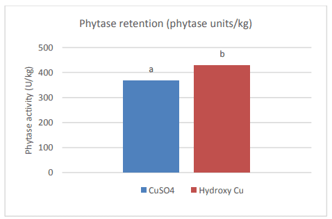
Graph 2: Effect of Cu sulphate and hydroxy Cu chloride on phytase retention. (a, b)Values with different letters are different (P < 0.05) from each other.

Next to Phytase, soluble Cu also has a negative effect on the fat content in feed. Dissociated, soluble Cu can initiate the lipid autoxidation cycle by playing a role as catalysator in the formation of free radicals, hereby reducing the nutritional value of the fat. A large difference was shown between the oxidation capacity of Cu sulfate and hydroxy Cu chloride (Miles et al., 1998). In this study, Feed grade Cu sulfate was sieved and coarse and fine Cu sulphates were retained with a respective modal particle size of 455 µm a b 0 100 200 300 400 500 Phytase activity (U/kg) Phytase retention (phytase units/kg) CuSO4 Hydroxy Cu Strong negative impact of sulphates No negative impact of TBCC Relative phytate P hydrolysis by phytase (%) and 11,9 µm. The two Cu sulphate fractions and hydroxy Cu chloride (modal particle size = 67 µm) were mixed in broiler diets at 25 ppm Cu. The peak peroxidation value observed for fine Cu sulphate was 5,8 times higher than for hydroxy Cu sulphate. At day 20, the anisidine value measured for fine Cu sulphate was 2,2 times higher than for hydroxy Cu sulphate. The values for coarse Cu sulphate were comparable to hydroxy Cu chloride at all time points and for both measured parameters. When using feed grade Cu sulphate, there will always be a part of fine size and this part will have a larger negative effect on lipid peroxidation compared to hydroxy Cu chloride. Although the average particle size of hydroxy Cu chloride was smaller than coarse Cu sulphate, the effect of oxidation was similar. Improved oxidative stability of soybean oil towards hydroxy trace minerals compared to sulphates was confirmed in an in vitro model using Rancimat Test (Kampf et al., 2012).

To assess the slow release effect of hydroxy Cu chloride throughout the GIT an in vivo study was conducted (Naziripour & Klasing, 2010). Broilers were fed 150 ppm Cu, from Cu sulphate and hydroxy Cu chloride, for 2 weeks (day 3 – 14). The amount of Cu (mg/kg DM) was analyzed and divided into water soluble Cu, EHPG extractable Cu and unremovable Cu (unavailable for nutritional and microbicidal purposes). Copper sulphate was shown to increase the duodenal luminal soluble Cu almost twofold compared to hydroxy Cu chloride, augmenting the negative effect of Cu on the villi and the tendency to form complexes with dietary nutrients resulting in a higher unremovable Cu part later on in the GIT. Hydroxy Cu chloride resulted in more EHGP-extractable Cu in all regions of the intestines (P < 0.05) and less unremovable Cu (P = 0.05), relating to its greater bioavailability and anti-coli activity. Hydroxy Cu chloride was shown to inhibit E.coli growth (P < 0.05) to a greater extent than Cu sulphate in the lower gastrointestinal tract. Performance-wise there is also a clear distinction between Cu sulphate and hydroxy Cu chloride (Kim & Kil, 2015) The inclusion of 100, 200 and 300 mg/kg added Cu from hydroxy Cu chloride resulted in an improved ADG, ADFI and FCR for all levels compared to Cu sulphate.

Hydroxy Zinc chloride has been researched in several animal species. Relative bio-availability was measured in piglets where improved bio-availability was shown for hydroxy zinc compared to zinc sulphate ranging from 122% (metacarpal zinc) to 159% (plasma zinc after 2 weeks of supplementation) (Zhang and Guo, 2007). In broilers, inclusion of 80 ppm hydroxy zinc chloride resulted in heavier birds and more breast meat yield compared to 80 ppm of zinc sulphate or a combination of 60 ppm zinc sulphate combined with 20 ppm zinc-chelate, without compromising feed conversion (Parr et al., 2013)

Selenium in feed – all about L-selenomethionine

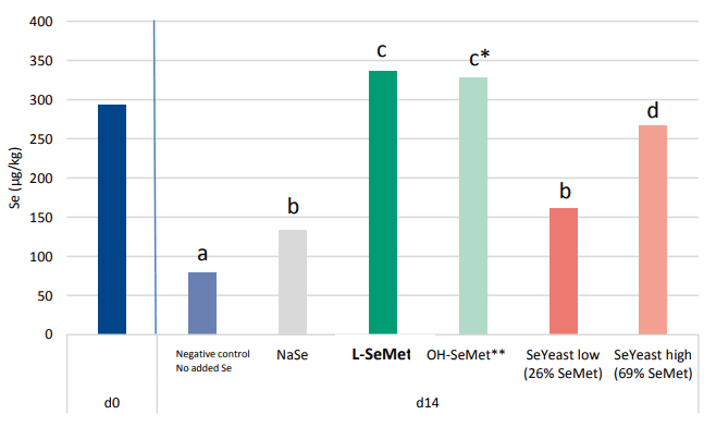
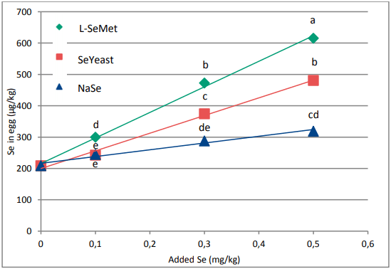
When looking at selenium, different sources are used in practice. While selenite was and still is used for a big extend in feeds, it is generally accepted that organic selenium is the best source of selenium. Organic selenium sources are evaluated on their content of L-Selenomethionine (L-SeMet). This unique form can be build up, in the same way as methionine, in the protein fraction of tissues and animal products (milk, eggs, meat). A study proved the enhanced Se deposition of L-SeMet compared to other selenium containing (in)organic sources in broiler breast meat (Graph 3; Van Beirendonck et al., 2016). Two Seyeasts were incorporated in the trial reflecting products that can be found in the market. The Se-yeast high in Se as L-SeMet (69% of the total Se content) showed a high deposition but significantly lower than the group fed L-SeMet. The Se-yeast low in L-SeMet (26%) showed even a significantly lower deposition compared to the Se-yeast high in L-SeMet. When plotting the Se deposition in function of the L-SeMet concentration, it can be clearly derived that the deposition is linearly proportional to the L-SeMet concentration in the product. OH-SeMet has a numerical lower deposition of Se in the muscle compared to L-SeMet. When the non-intended overdose is taken into account, the selenium deposition confirmsthe efficacy of 80% for the conversion of OH-SeMet to L-SeMet in the animal, as described for OH-Methionine (EFSA Journal 2012;10(3):2623).

Graph 3: Se deposition in broiler muscle (µg/kg) at day 14 after supplementation of different Se sources (total added Se = 0.2 mg/kg); (a-d)Values with no common superscript, differ significantly (P < 0.0001); * Result of a non-intended overdose of 14% due to a difference between spec value and the actual measured total Se value of the product. Total added Se = 0.228 mg/kg; ** OH-SeMet does not contain L-SeMet. OH-SeMet has to be first converted into L-SeMet in the body. Conversion efficiency is 80% (EFSA Journal 2012;10(3):2623).

Next to the deposition in meat, the most efficient transfer of Se to eggs can also be achieved by adding LSeMet (Graph 4; Delezie et al., 2014). The study proved that the addition of L-SeMet is superior to Seyeast and sodium selenite in augmenting the total Se deposition in eggs. The superior deposition of LSeMet compared to the other sources ensures an optimal delivery to the animal and offspring, by increasing the Se pool, and a reduction in oxidative stress (e.g. high growth, early life stages). Stress was reduced significantly (P < 0.05) in chick embryos by increasing the selenium pool via L-SeMet in broiler breeders (Xiao et al., 2016).

Graph 4: Se deposition in eggs (µg/kg) after 56 days of treatment. (a-d)Values with no common superscript, differ significantly (P < 0.05)

Organic selenium has many practical applications in poultry diets (Table 2; Fisinin et al., 2008). Next to improved performance parameters, selenium may improve fertility, anti-oxidant status, meat characteristics and carry-over of selenium to offspring or meat and eggs for human nutrition.

Table 1: Practical applications of organic selenium in poultry diets. Fisinin et al., 2008; Current Advances in Selenium Research and Applications. Volume 1. Page 221 – 261

Feed for thought for formulators

With regard to Cu and Zn, extensive data showed the advantages of using hydroxy trace chlorides in broiler and layer feed. Advantages can be seen as well in the feed as in the animal and its performances. Focusing on selenium, choosing L-SeMet is the way to go. Enhancement of the selenium pool of the animals cannot only provide benefit to the animal itself but also its progeny and the consumers relying on the products derived.

References available on request

Bruneel@orffa.com

Eising@orffa.com